 Skip to content
Ophthalmology CRO
Ichor Life Sciences is an Ophthalmology CRO with over a decade of experience helping researchers move promising therapies toward the clinic. Our preclinical ophthalmology programs are designed with clinical translation in mind, helping sponsors generate robust data to guide development decisions.

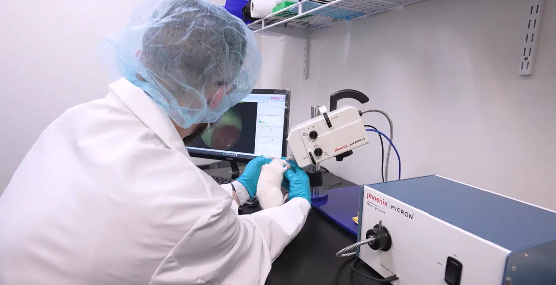
Strong ophthalmic programs begin with preclinical studies designed around the questions that matter in the clinic. As an experienced Ophthalmology CRO, Ichor Life Sciences supports ocular drug development with relevant disease models, specialized endpoints, and study strategies built to inform the path forward. Our team brings more than a decade of ophthalmology experience to every program.
Translational Ophthalmology CRO Capabilities
As an experienced Ophthalmology CRO, Ichor Life Sciences combines specialized ocular models with endpoints that help researchers understand how a therapy is performing before entering the clinic. Our approach considers the structural, functional, and biological measures that can help connect preclinical findings with future clinical development.
Ocular Function and ERG Assessment
Understand how disease and treatment affect retinal function with electroretinography ("ERG"). ERG provides functional measurements that complement anatomical findings and can help establish a clearer picture of treatment effect before clinical evaluation.
Ocular Dosing and Targeted Delivery
Route of administration can play an important role in ophthalmic drug development. Our team performs intravitreal, subretinal, intracameral, suprachoroidal, and topical dosing using specialized systems suited for precise delivery to the eye.
Longitudinal Retinal Imaging with SD-OCT
Follow structural changes in the retina over the course of a study using spectral-domain optical coherence tomography ("OCT"). SD-OCT provides high-resolution measurements that allow investigators to monitor disease progression and treatment-related changes over time.
IOP Monitoring for Glaucoma Studies
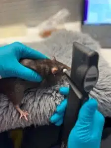
Reliable intraocular pressure measurements require careful control of factors that can influence the result. Animals are acclimated through positive reinforcement training to reduce handling-related stress and improve consistency during repeated IOP measurements.
Ocular Histology and Tissue Analysis
Investigate what is happening at the tissue and molecular level through specialized ocular collection and analysis. Our capabilities include retinal flat mounts, whole-eye collection, H&E, IHC, IF, flow cytometry, ELISA, western blotting, and mass spectrometry.
Retinal Vascular and Autofluorescence Imaging
Visualize changes in retinal vasculature with fluorescein angiography and assess additional ocular features through fundus autofluorescence. Quantitative image analysis can be incorporated to compare findings across treatment groups and study timepoints.
Fundus Imaging and Quantitative Readouts
Capture detailed views of ocular structures with high-definition fundus imaging. Automated image analysis provides quantitative measurements that can strengthen interpretation of disease progression and treatment effects while supporting endpoints relevant to clinical development.
Endpoints with Goals in Mind

As an experienced Ophthalmology CRO, we use advanced ocular imaging to produce clinically relevant endpoints. This mindset allows us to provide a clearer view of treatment effects and help inform decisions as programs move toward the clinic.
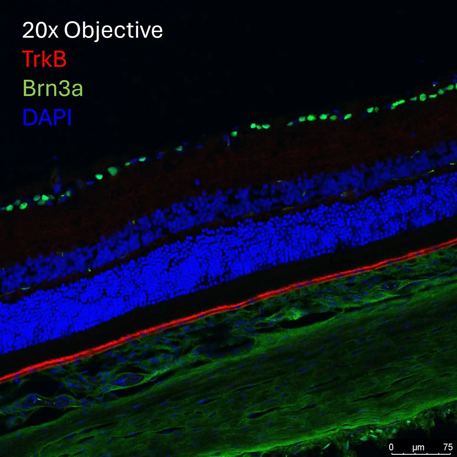
Efficacy is only part of the story. Understanding target engagement and mechanism of action can provide the biological context behind treatment effects and strengthen the evidence supporting progression toward clinical studies.
Capabilities and Services Summary
Facilities and Animal Welfare
Facilities and Animal Wellness
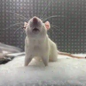
Our vivarium is an AAALAC accredited facility that supports rapid protocol review. We are an independent company with 100% of our operations in the United States. Consistent with a strong ethos towards animal welfare, our positive reinforcement training (PRT) program and enrichment standards are world class and industry-leading.
Equipment
Equipment
Ichor uses the most state-of-the-art equipment in the field of ophthalmology. This includes the Leica spectral-domain optical coherence tomography (OCT), Phoenix Micron IV, TonoPen and TonoLab, and World Precision Instrument’s microinfusion pump system for intraocular drug delivery. The advantage of using the Leica OCT allows for more accurate scans in rodent models, precise retinal thickness measurements, and high-quality resolution images of retinal architecture. With the Phoenix Micron IV, we can perform fundoscopy, fundus autofluorescence, fluorescein angiography, anterior imaging, blue light illumination exposure. In combination with our IOP readings with our tonometers we implement the use of positive reinforcement training to allow for conscious-anesthesia free IOP measurements. Full field ERGs are also available upon request. The use of WPI’s microinfusion injection equipment allows for accurate and reproducible intraocular injection procedures allowing us to complete intravitreal, subretinal, intracameral, and suprachoroidal injections.
We Are Always Evolving
We Are Always Evolving
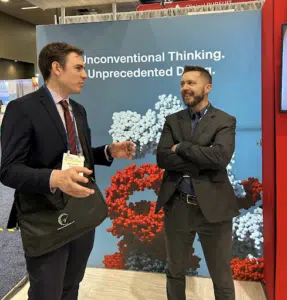
Our ophthalmic cro team members regularly attend scientific conferences to keep up to date with the latest models, instrumentation, and therapeutic targets. We partner with technology companies developing next generation analytical instrumentation and biomarkers to always be on the forefront of industrial trends and client needs.