 Skip to content
Ophthalmic CRO
Ichor Life Sciences is a premier ophthalmic cro with over a decade of experience tackling the toughest challenges in ophthalmic disease research. Discover why we are the fastest growing preferred partner in ophthalmology.

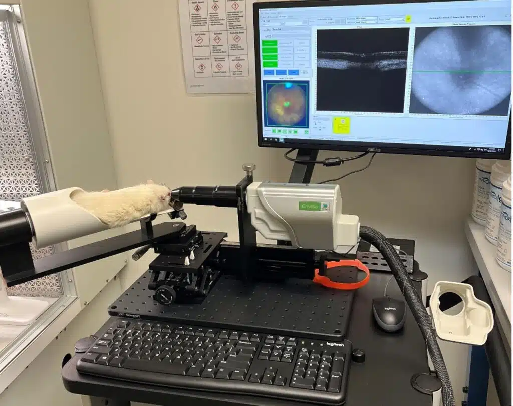
Spanning anterior to posterior disease models, Ichor Life Sciences is an established ophthalmic cro providing preclinical research services to support the development of innovative ophthalmic therapies. With over 10 years of experience in space, we have seen and supported some of the most diverse models. Let our scientific staff assist you with developing your investigational product today!
Featured Capabilities
Our ophthalmic cro research team utilizes industry-best equipment to conduct studies. Quality equipment for quality results.
Retinal scans using spectral-domain optical coherence tomography
Track retinal disease progression using superior image quality with SD-OCT. More accurate measurements means clearer characterization of pathogenesis in our disease models.
High-definition fundoscopy with automated image quantification
Obtain high definition images in anterior and posterior segment disease models to better inform pathogenesis and therapeutic response. Our automated scoring algorithms provide robust, quantitative read-outs.
Advanced intraocular drug delivery capabilities
Microinfusion pumps and micromanipulator devices for rodents provide accurate, efficient, and proficient drug delivery. Available administration modalities include intravitreal, subretinal, intracameral, suprachoroidal, and topical.
Complex dissections, bioanalysis, and histopathology capabilities
We perform complex dissection for all ocular tissues. From flat-mounts to whole eye collections, we provide H&E, IHC, and IF processing, flow cytometry, ELISA, western-blot, and mass spectrometry.
Advanced retinal function testing with electroretinography
Evaluate the preservation or restoration of ocular function with high quality ERGs. This powerful technique tests retinal function in response to electrical impulses.
High-definition fluorescein angiography
We provide clear and consistent fluorescent angiograms and fundus autofluorescence measurements for a variety of different ocular models. Quantitation using automated scoring is also available.
Industry leading intraocular pressure measurements
IOP measurements can be compromised by external effects, leading to false readings. Our positive reinforcement training (PRT) ensures study animals are stress free to provide reliability and consistency in our measurements.
Your Results Await
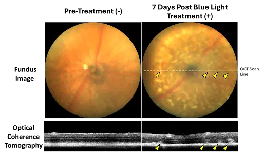
This featured Stargardt disease research model utilizes the ABCA4-/- transgenic knockout mouse for testing gene therapy and other investigational products for Stargardt disease and dry age-related macular degeneration. Blue light illumination in the ABCA4-/- strain activates reactive lipofuscin fluorophores and induces retinal lesions, which can be measured by fundus imaging, OCT, and histology. Functional impairment by ERG is also observed. This model is ideal for interventions aimed at directly or indirectly reducing lipofuscin burden or cytoprotective interventions intended to preserve RPE and photoreceptor structure and function.
Endpoints with Goals in Mind
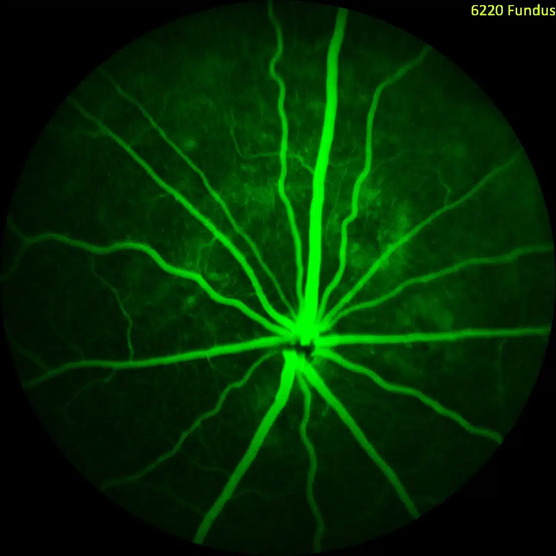
Advanced Imaging in CNV Model
We are an ophthalmic cro that utilizes state-of-the-art imaging modalities to provide the most robust data sets in support of client projects. If a picture is worth 1,000 words our data speaks volumes.
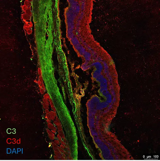
Histology for Target Engagement
We do not stop just at efficacy endpoints. Confirmation of target engagement and a clear understanding of mechanism of action inform downstream product development.
Capabilities and Services Summary
Facilities and Animal Welfare
Facilities and Animal Wellness
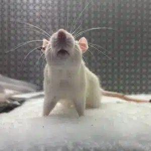
Our vivarium is an AAALAC accredited facility that supports rapid protocol review. We are an independent company with 100% of our operations in the United States. Consistent with a strong ethos towards animal welfare, our positive reinforcement training (PRT) program and enrichment standards are world class and industry-leading.
Equipment
Equipment
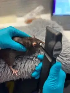
Ichor uses the most state-of-the-art equipment in the field of ophthalmology. This includes the Leica spectral-domain optical coherence tomography (OCT), Phoenix Micron IV, TonoPen and TonoLab, and World Precision Instrument’s microinfusion pump system for intraocular drug delivery. The advantage of using the Leica OCT allows for more accurate scans in rodent models, precise retinal thickness measurements, and high-quality resolution images of retinal architecture. With the Phoenix Micron IV, we can perform fundoscopy, fundus autofluorescence, fluorescein angiography, anterior imaging, blue light illumination exposure. In combination with our IOP readings with our tonometers we implement the use of positive reinforcement training to allow for conscious-anesthesia free IOP measurements. Full field ERGs are also available upon request. The use of WPI’s microinfusion injection equipment allows for accurate and reproducible intraocular injection procedures allowing us to complete intravitreal, subretinal, intracameral, and suprachoroidal injections.
An Image Worth 1,000 Words
An Image Worth 1,000 Words
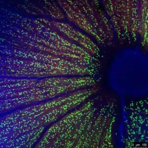
We understand that decisions are made every day based on the data we produce, and we recognize the responsibility to deliver on our clients’ trust in us. Whether it be OCT scans to inform a winner amongst multiple lead candidates, or vibrant histology slides stained in your company’s colors to wow investors, we are firmly committed to providing the highest quality data possible for your needs.
We Are Always Evolving
We Are Always Evolving
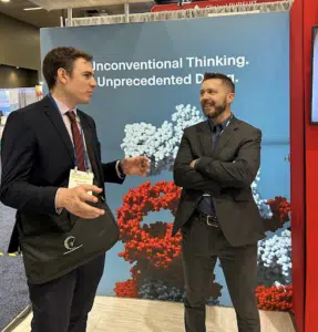
Our ophthalmic cro team members regularly attend scientific conferences to keep up to date with the latest models, instrumentation, and therapeutic targets. We partner with technology companies developing next generation analytical instrumentation and biomarkers to always be on the forefront of industrial trends and client needs.
Frequently Asked Questions
Answers to some of the most common questions about our ophthalmic cro service offerings.