 Skip to content
Glaucoma Models
Leverage our experience and proficiency in glaucoma animal models to meet the development needs of your investigational products and research programs.

Choose from our validated pre-clinical glaucoma animal models spanning optic nerve crush, ocular hypertension, and more. Need custom services? Our skilled team can customize or onboard new models to meet diverse project needs.
Featured Capabilities
Our team of ophthalmic experts has supported research with glaucoma animal models for decades. Learn more about our capabilities.
Trust Our Team of Experts

Our interdisciplinary team of skilled husbandry, ophthalmic surgical, and analytical staff offer several glaucoma models which can be customized to meet your specific project needs and budget. We continue to invest heavily in the development and onboarding of new glaucoma animal models to support the needs of our clients and collaborators.
Best-in-class Animal Handling Practices

Positive Reinforcement Training Improves Data
Ichor is pioneering best-in-industry animal handling practices to reduce stress and improve data, which includes PRT as standard offering in many of our glaucoma models.

Glaucoma Animal Model Customization
We understand your project needs may extend beyond the norm. Our scientific team will work with you to customize current and new service offerings to meet your needs.
Featured Case Studies / Capabilities
Optic Nerve Crush Glaucoma Animal Model
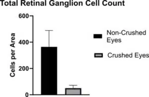
Optic Nerve Crush Glaucoma Animal Model
We provide the ability to track disease progression after surgical induction by utilizing optical coherence tomography (OCT), measuring full retinal thickness, the retinal ganglion cell (RGC) layer and nerve fiber layer (RNFL) in real time. Post-life retinal flat mounts and histological sections can quantify retinal ganglion cell loss (shown) to complement structural measurements.
Laser-Induced Ocular Hypertension Model
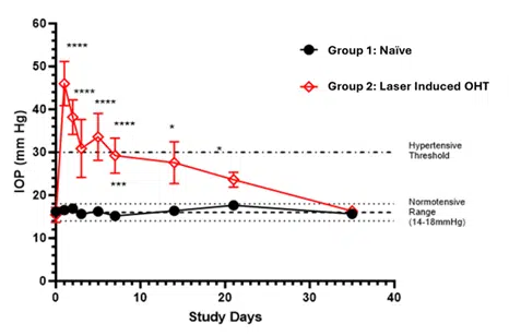
Laser-Induced Ocular Hypertension Model
Laser ablation results in sustained increase in intraocular pressure for several weeks duration, making this a robust and preferred glaucoma model for many clients.
Intraocular Pressure Glaucoma Model
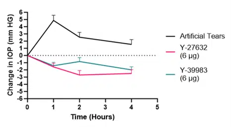
Intraocular Pressure Glaucoma Model
Rodent intraocular pressure models feature rapid and robust assessment of investigational products. Select from Latanoprost or rho-kinase (ROCK) controls based on your mechanism of action.