 Skip to content
In Vitro Angiogenesis Assays
Angiogenesis is a dynamic and intricate biological process with far-reaching roles in health and disease. Ichor’s 2D and 3D cell-based angiogenesis assay portfolio quantifies pro- and anti-angiogenic activity through live-cell kinetic imaging.

What Can You Measure?
Build a more complete angiogenesis profile by measuring network formation, migration, and sprouting across integrated 2D and 3D in non-invasive angiogenesis assays.
| Assay | Function | Key Readouts |
|---|---|---|
|
2D Tube Formation Assay
|
Quantifies how endothelial cells assemble into interconnected vascular-like networks, revealing treatment effects on network extent, branching, connectivity, and organization over time. |
Total length: measures the overall extent of vascular-like network formation.
Master junctions: reflect network maturity and interconnected complexity.
Branching intervals: capture branching activity across the developing network.
Mesh index: characterizes network organization and maturation.
|
|
2D Scratch Wound / Migration Assay
|
Tracks endothelial migration and wound closure kinetics in real time, revealing how treatment alters cell movement during the angiogenic response. |
Relative wound density: measures the degree of endothelial migration into the wound area.
Wound-closure kinetics: tracks the progression of endothelial migration and closure over time.
Angiogenesis-related cytokines: characterize signaling associated with the broader angiogenic response.
|
|
3D Spheroid Sprouting Assay
|
Measures endothelial sprouting from HUVEC spheroids in a three-dimensional matrix, capturing a spatially complex angiogenic phenotype alongside the 2D assays. |
Total spheroid sprout length: measures the extent of endothelial sprout outgrowth into the surrounding 3D matrix.
|
Angiogenesis Assays in Aging Research

As research into aging and age-related disease gains momentum, angiogenesis assays offer a powerful functional window into vascular health and regenerative capacity.
Featured Case Studies / Capabilities
Tube Formation Assay
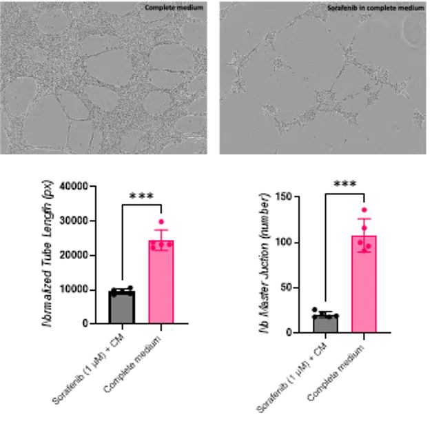
The HUVEC tube formation assay offers a fast, visually compelling way to see angiogenic activity take shape in real time. Using live-cell kinetic imaging, we follow the development of endothelial networks and quantifies features such as tube length, junctions, and meshes to capture how a test article influences vascular organization and connectivity.
Because tube formation represents only one dimension of angiogenesis and can show inherent variability, Ichor pairs it with endothelial migration and 3D spheroid sprouting assays to build a stronger understanding of how a test article modulates the angiogenic response.
Scratch-Wound Assay
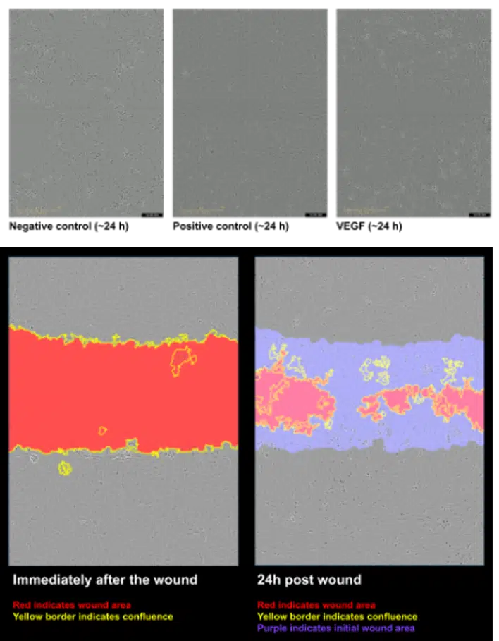
Endothelial migration is a critical step in angiogenesis, enabling cells to move toward angiogenic signals and contribute to new vessel growth. The scratch-wound assay follows this process in real time, using live-cell imaging and relative wound density to capture treatment-driven changes in migration and wound-closure kinetics.
To add another layer of biological insight, terminal supernatant can be analyzed using an angiogenesis-focused cytokine bead array, connecting changes in cell behavior with key angiogenic signaling factors. Time-lapse movies can also provide a compelling visualization of the response as it unfolds.
Spheroid Sprouting Assay
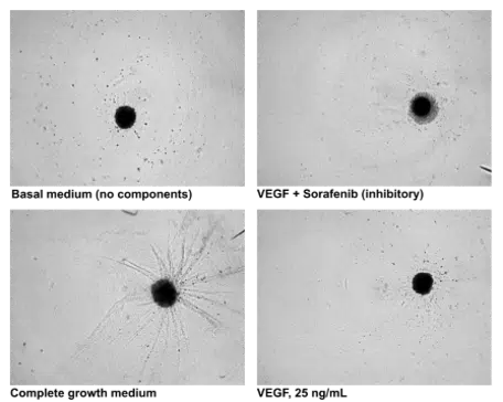
Endothelial sprouting is a key step in new vessel growth, extending beyond the flat-cell behaviors captured in 2D assays. The 3D spheroid sprouting assay follows HUVEC sprout outgrowth within a matrix using live-cell imaging, providing a more spatially complex view of the angiogenic response.
Custom image analysis quantifies total spheroid sprout length, while representative condition images show how the response changes across treatments. This 3D readout adds a distinct layer of functional insight alongside tube formation and endothelial migration.
Frequently Asked Questions
Need more information about our angiogenesis assays? Learn more below, or reach out to our team with any questions.