 Skip to content
Cell Viability Assays
We offer an extensive collection of cell viability assays to support your studies. Our facilities are equipped to conduct high-throughput microplate assays with kinetic or end-point readouts.

In addition to standard cell proliferation assays, we can create custom cell-based assays based on your needs. UV/Vis, fluorescent, and luminescent assays can be tailored to answer your research questions.
Cell Viability Assays
Cellular respiration is assessed by tetrazolium reduction as a reliable reporter of cellular viability and metabolic activity.
Cell Proliferation Assays
To evaluate cell proliferation, nucleotide analog incorporation can be assessed by downstream click chemistry with a customizable fluorophore.
Cellular Apoptosis Assays
An established cell apoptosis assay uses phosphatidylserine display, an early hallmark of apoptosis, to measure apoptosis initiation via a fluorescent reporter.
Reactive Oxygen Species Assays
Presence of reactive oxygen species (ROS) is measured by addition of a probe that fluoresces upon reaction with intracellular ROS.
Aggresome Formation Assays
A cell-based assay for aggresome formation reflects the cellular response to protein misfolding.
State-Of-The-Art Instrumentation Enables Discovery
Our advanced instrumentation and reliable assays deliver trusted results.
Your Toolkit for Cell Viability

Cell death can proceed by different pathways, and important events are missed when observations are limited by assay availability. Our collection of cell viability assays can differentiate between apoptosis, necrosis, and senescence. We also provide several methods of analysis, including plate reads, microscopy, and flow cytometry. We have the tools to deliver definitive results.
Complementary Assays to Characterize Cell Death

Flexible Fluorophores with Click-iT EdU
For experiments with multiple fluorophores, Click-iT EdU provides options for click-chemistry-enabled labeling, avoiding spectral overlap when multiple fluorophores are used.

Annexin-V Discriminates Between Apoptosis and Necrosis
An advantage of staining with Annexin-V FITC is that it is specific for apoptosis, providing information on the mechanism of cell death.
Featured Case Studies / Capabilities
CellTiter AQueous
Cellular Metabolism and Viability
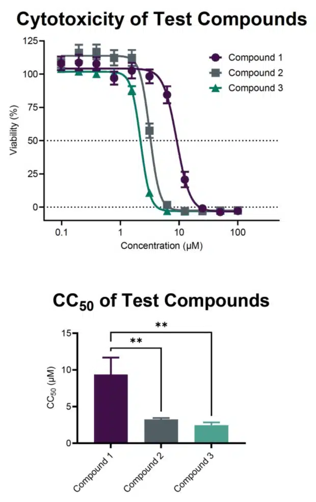
The Promega CellTiter AQueous Cell Proliferation Assay resembles the canonical MTT assay. As cells generate NADH as a consequence of metabolism, the assay substrate, a tetrazolium salt, is reduced by NADH to form formazan. Formazan production can be measured by absorbance at 490 nm to quantitate cellular viability. An important advantage of the CellTiter AQueous method is that it avoids a cell lysis step prior to endpoint analysis. This enables kinetic reads to measure changes in cell viability or metabolism in real time.
Here, transformed cells were exposed to concentration gradients of test compounds, and cell viability was calculated as an endpoint by the CellTiter Aqueous cell viability assay. Percent viability was calculated relative to vehicle, and CC50 values were calculated from the curves. This method demonstrates significant differences in CC50 values for select test compounds.
Click-iT EdU
Cellular Proliferation
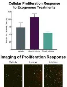
The Invitrogen Click-iT EdU Cell Proliferation Assay is a modification of the classic BrdU incorporation assay. In contrast to the BrdU assay, Click-iT EdU utilizes an unlabeled nucleotide analogue for incorporation into newly synthesized DNA, then takes advantage of click chemistry to append a fluorescent conjugate of choice to the incorporated analogue for end-point analysis. The benefit of this approach is that a variety of fluorophore options are available, thereby accommodating existing staining and imaging workflows.
Here, cell proliferation in various growth conditions was measured by the Click-iT EdU cell proliferation assay. Cells were grown with growth stimulator, growth repressor, or vehicle control. Cell proliferation was measured as the percent EdU positive cells. Significant differences were observed for either treatment versus media alone.
Click-iT EdU can be used for endpoint proliferation measurement, pulse-labeling, or cell imaging applications.
Annexin V-FITC

Apoptosis Assay
Annexin V staining is a well-characterized method for quantitating cellular apoptosis. The Enzo Annexin V-FITC apoptosis assay detects cells as they initiate the apoptotic cascade by virtue of their phosphatidylserine display. This early hallmark of programmed cell death can be measured by Annexin V staining, due to the high affinity and specificity of Annexin V for phosphatidylserine. Because Annexin V cannot pass through the cell membrane, only apoptotic cell displaying phosphatidylserine in the outer membrane leaflet will stain positive with Annexin V conjugates.
Here, cells were incubated with increasing concentrations of apoptosis-inducing treatment. After 24 hr, the cells were assessed for apoptosis using the Annexin-V FITC method. Apoptosis was calculated as the ratio of fluorescent intensity to number of nuclei (stained with Hoechst). A dose-dependent increase in Annexin-V FITC staining was observed. Cells were also imaged at experimental endpoint.
Annexin-V FITC stained cells can also be analyzed by flow cytometry.
PROTEOSTAT

Aggresome Detection
The Enzo PROTEOSTAT Aggresome detection kit provides a cell-based assay for intracellular aggresome formation. Cells sequester misfolded proteins to form aggresomes as a defense mechanism. The formation of aggresomes is an active and regulated process to protect against the deleterious effects of aberrantly folded proteins. Monitoring aggresome formation provides information about proteostasis, and interventions targeting aggresome formation can alter the cell’s ability to mitigate misfolded proteins.
Here, cells were treated with various concentrations of a known inducer of aggresomes, then stained for aggresome presence. The quantity of aggresomes per field of view was measured, and demonstrated a dose-dependent increase with increasing treatment by aggresome inducer. Cell images corroborate graphed results.
ROS-ID
Reactive Oxygen and Nitrogen Detection
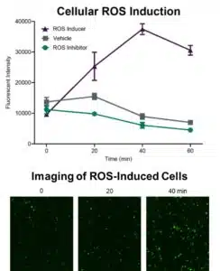
The Enzo ROS-ID Total ROS detection kit measures intracellular reactive oxygen species (ROS) and reactive nitrogen species (RNS). ROS and RNS are implicated in cell signaling and homeostasis, but can also impart deleterious effects when dysregulated. ROS-ID detects both ROS and RNS via a small molecule probe that permeates the cell membrane, then fluoresces upon reaction with ROS or RNS. The assay can be conducted in live cells, and results can be measured by fluorescent plate reads, fluorescent cell imaging, or flow cytometry.
Here, cells were treated with either an ROS inducer, an ROS inhibitor, or vehicle control, and fluorescence was measured over time by plate reads. An increase in ROS/RNS over time was observed for cells treated with ROS inducer, peaking at 40 min. ROS inhibitor led to fluorescent values below those of vehicle-treated cells. Imaging of the cells treated with inducer also show increased fluorescent signal over time.
Frequently Asked Questions
Read more to find out which assay is best for your project.