 Skip to content
Endotoxin Induced Uveitis (EIU) Model
Our unique endotoxin induced uveitis model produces definitive, repeatable disease characteristics. By administering endotoxin via intravitreal injection, our model circumvents systemic response, and more accurately reflects the clinical disease state.

Endotoxin induced uveitis is graded by anterior imaging, assessing disease progression over time. In vivo monitoring of clinical signs provides more opportunities to measure the effects of interventions compared to endpoint analyses alone.
Intravitreal Endotoxin Injection
Endotoxin is administered locally, avoiding systemic effects and mimicking human disease.
Pronounced, Reproducible Disease Characteristics
Well-defined disease state and low model variability yield a sensitive system for measuring interventions.
Customized Treatment and Readout Capabilities
Our veterinary team can incorporate treatment methods to suit your research needs, and create customized endpoints for your study.
Cytokine Measurement
Inflammatory markers are assessed from aqueous humor and vitreous humor for disease context.
Histopathology Scoring
H&E staining and reading is conducted to provide quantitative scores of retinal, anterior chamber, and ciliary body pathology.
Superior and Customizable EIU Studies
Ichor’s full-service research capabilities extend to our endotoxin induced uveitis model.
Intravitreal injections are conducted by our skilled and experienced veterinary team, using fundoscope-guided delivery. This ensures precise administration of endotoxin and guarantees a robust, repeatable disease state.
Tissue sections are prepared and read by our board-certified pathologist to evaluate tissue architecture and immune cell infiltrate as measures of inflammation and disease state.
We can incorporate your desired endpoints into our existing EIU model. Integrating readouts prevents the need for additional animal studies, and provides paired endpoints for more consistent data analysis.
Our veterinarian conducts clinical assessment of EIU disease in-house, providing rapid assessment of disease progression and efficacy of interventions.
Aqueous humor and vitreous humor are collected and analyzed for cytokines and chemokines to evaluate local inflammatory environments. Panels can be customized to meet your needs.
In Vivo Imaging Monitors Disease Progression

Standard experimental endpoints don’t provide information about the temporal aspects of disease progression. By imaging the anterior segment for clinical pathology throughout the course of the experiment, we can capture dynamic differences in endotoxin induced uveitis severity that are not reflected by endpoint analysis alone. This clinical assessment is sensitive enough to detect significant differences between healthy (untreated), EIU, and EIU + intervention groups.
Pronounced Pathology Means Significant Results

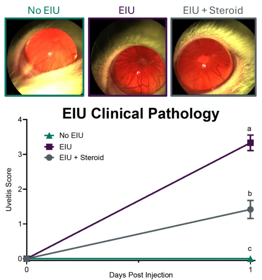
In vivo imaging of anterior segment
Endotoxin induced uveitis model shows significant differences in clinical disease signs between healthy (untreated), EIU, and EIU + intervention groups.

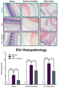
Histopathology of various eye tissues
Endpoint histopathology scores are significantly different between No EIU, EIU, and EIU + Steroid groups for all three tissues analyzed.
Featured Case Studies / Capabilities
In Vivo Imaging
Anterior Segment Imaging for Clinical Signs of Disease
In vivo imaging is conducted to monitor endotoxin induced uveitis disease progression. Anterior segment images are scored to grade disease severity. By two-days post injection, endotoxin induced uveitis is evident in endotoxin-treated groups. Disease state is consistent, producing significant clinical signs and minimal error, and allowing for sensitive assessment of interventions. Here, steroid treatment shows significant reduction in uveitis score. The reproducibility and extent of disease yield a sensitive model and make interventional effects obvious.
Histopathology
H&E Staining and Scoring for Pathology Assessment
Tissues are sectioned and H&E stained, and scored for pathology as an experimental endpoint. Retinal layers, anterior chamber, and ciliary body sections are routinely performed. Endotoxin induced uveitis elicits significant pathology in each tissue, with reproducible disease severity. Here, steroid treatment shows a significant reduction in EIU pathology, demonstrating the sensitivity of the model to experimental interventions.
Frequently Asked Questions
There are several advantages to conducting your EIU experiment with Ichor.
We use fundoscope-guided intravitreal injection to induce EIU, providing a more relevant model. We also offer live imaging, histopathology, and cytokine analysis readouts, in addition to custom endpoints as requested.
By delivering endotoxin directly to the target site, intravitreal injection elicits more robust inflammation, resulting in a more pronounced disease state. Additionally, the localized response is more reflective of human disease.
Our skilled veterinary team can tailor drug administration to your needs. We routinely perform system and local administration, including topical administration or fundoscope-guided intraocular injection.
By injecting endotoxin intravitreally, our endotoxin induced uveitis model shows disease onset within hours, and experimental endpoints are collected 24 hours post-induction.