 Skip to content
Dry-Eye Disease (DED) Model
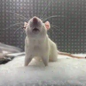
Dry eye disease (DED) is a prevalent ocular disorder characterized by disruption of the tear film that protects and lubricates the corneal surface. Our dry eye disease model combines controlled environmental conditions to replicate primary clinical features of the ocular surface disease.

To model evaporative stress, animals are housed in a Biospherix environmental chamber with tightly regulated conditions. Relative humidity is reduced to induce ocular surface drying, while temperature and airflow are controlled. The chamber creates a uniform low-humidity environment that increases tear evaporation in a physiologically relevant manner. Under these conditions, the ocular surface experiences progressive desiccation, leading to disruption of the tear film and epithelial stress
Controlled Environment
Low-humidity housing induces physiologically relevant evaporative stress without direct airflow to the face.
Tear Suppression
Scopolamine inhibits lacrimal secretion, producing sustained aqueous tear deficiency.
Ocular Damage
Progressive tear film instability leads to corneal and conjunctival epithelial disruption.
Inflammation
Desiccating stress activates inflammatory signaling pathways at the ocular surface.
Translational
Model recapitulates hallmark features of human dry eye disease.
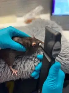
Featured Capabilities
Our preclinical ophthalmic research team utilizes industry-best equipment to conduct studies. Quality equipment for quality results.
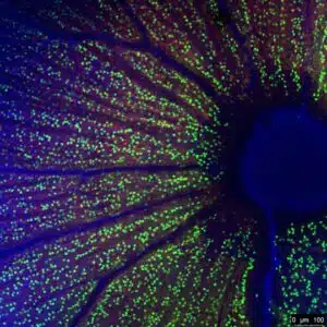
Representative Data

Objective assessment of dry eye severity is available through automated corneal image quantification. Following fluorescein staining, high-resolution corneal images are analyzed using a custom Python-based script to calculate mean fluorescence intensity across the corneal surface. This automated approach provides a reproducible, non-subjective readout of corneal fluorescein uptake, enabling quantitative comparisons across treatment groups and supporting robust efficacy assessment in dry eye studies.

Dry eye is evaluated using the NEI Density grading system, a widely accepted subjective scoring method that assesses punctate corneal fluorescein staining across five anatomical regions: nasal, temporal, inferior, superior, and central cornea. Each region is graded on a 0–3 severity scale, with scoring performed by a trained examiner masked to treatment groups. This approach enables sensitive detection of regional corneal damage and is well suited for efficacy comparisons in therapeutic intervention studies.
Capabilities and Services Summary
Facilities and Animal Welfare
Facilities and Animal Wellness
Our vivarium is an AAALAC accredited facility that supports rapid protocol review. We are an independent company with 100% of our operations in the United States. Consistent with a strong ethos towards animal welfare, our positive reinforcement training (PRT) program and enrichment standards are world class and industry-leading.
Equipment
Equipment
Ichor uses the most state-of-the-art equipment in the field of ophthalmology. This includes the Leica spectral-domain optical coherence tomography (OCT), Phoenix Micron IV, TonoPen and TonoLab, and World Precision Instrument’s microinfusion pump system for intraocular drug delivery. The advantage of using the Leica OCT allows for more accurate scans in rodent models, precise retinal thickness measurements, and high-quality resolution images of retinal architecture. With the Phoenix Micron IV, we can perform fundoscopy, fundus autofluorescence, fluorescein angiography, anterior imaging, blue light illumination exposure. In combination with our IOP readings with our tonometers we implement the use of positive reinforcement training to allow for conscious-anesthesia free IOP measurements. Full field ERGs are also available upon request. The use of WPI’s microinfusion injection equipment allows for accurate and reproducible intraocular injection procedures allowing us to complete intravitreal, subretinal, intracameral, and suprachoroidal injections.
An Image Worth 1,000 Words
An Image Worth 1,000 Words
We understand that decisions are made every day based on the data we produce, and we recognize the responsibility to deliver on our clients’ trust in us. Whether it be OCT scans to inform a winner amongst multiple lead candidates, or vibrant histology slides stained in your company’s colors to wow investors, we are firmly committed to providing the highest quality data possible for your needs.
We Are Always Evolving
We Are Always Evolving
Our preclinical ophthalmic research team members regularly attend scientific conferences to keep up to date with the latest models, instrumentation, and therapeutic targets. We partner with technology companies developing next generation analytical instrumentation and biomarkers to always be on the forefront of industrial trends and client needs.