 Skip to content
Cell Painting Assay
Cell painting combines high content imaging with multiplexed fluorescent labels to deliver incredibly detailed cell microscopy. This technique provides information on cell composition, morphology, and trafficking.

Ichor’s cell painting assays provide detailed, high resolution images to characterize cellular organization and composition, along with automated workflows for analyte characterization and quantitation.
Receptor Internalization and Trafficking
Cell painting facilitates the tracking of cellular receptors. Differential permeabilization provides information on receptor surface presentation or internalization.
Multiplex Labeling
Ichor’s cell painting assays can incorporate up to five fluorescent labels in a single imaging workflow. The simultaneous labeling of multiple cell features provides more information on cell architecture and composition.
Colocalization Analyses
Like traditional confocal microscopy, cell painting assays can be used to infer analyte colocalization, but cell painting is uniquely capable of the simultaneous interrogation and overlay of several cell features.
Organelle Tracking
Multiplex labeling can characterize organelle abundance and distribution, while co-localization analyses demonstrate organelle trafficking and fusion events.
Cell Maturation and Differentiation
As cells differentiate, their morphology and composition changes. Cell painting captures these changes in high resolution, and quantitates maturation features.
Versatile Instruments and Approaches
We use a suite of cell imaging instruments and offer customized workflows to provide superior cell painting for every project.
Advanced software performs automated analyses, utilizing machine learning technology to answer complex cell imaging queries. Image collection and sophisticated analyses deliver precise and reliable results.
Select assays can be performed on live cells during time course experiments to observe temporal trends. For other experiments, sampling and fixing cells at designated time points enables characterization of dynamic cell responses.
For streamlined experiments, Ichor will perform cell or animal treatments in-house and process samples for cell painting assays. Integrating in vivo experiments with downstream imaging improves quality, efficiency, and reproducibility.
Take advantage of our pre-established labeling methods for common cell features, and incorporate your own stains or antibodies for custom labeling profiles. We work with you to optimize label compatibility.
Automated field-of-view sampling collects quantitative data from confocal images. This method removes sampling bias, and provides additional data points for more precise quantitation.
Automated Image Analysis and Quantitation

Ichor cell painting procedures enable the imaging of live or fixed cells, 2D or 3D image acquisition, multiplex cell painting with a wide assortment of fluorophores, and high-resolution images acquired by spinning disc confocal microscopy. Additionally, automated workflow and calculation protocols provided reliable quantitation of cell painting assays. Our high content imagers use state-or-the-art processing algorithms to define regions of interest based on fluorophore distribution. This allows for identification of subcellular compartments and organelles, and for the specific interrogation of probe intensity in designated areas. Additionally, automated field of view sampling minimizes bias in measurements and improves the accuracy of your results.
An Image Worth 1,000 Words

High Resolution Multiplex Labeling
The use of multiple probes in cell painting assays provides high-resolution characterization of cell structure, morphology, and composition. Distribution or co-localization of cellular features can be assessed by this method.

3D Imaging for Spheroid Characterization
Ichor’s cell painting capabilities include characterization of 3 dimensional (3D) assemblies. Our analysis software features an XYZ-viewer to assess structural and compositional attributes in 3D spheroids, organoids, or tissue explants.
Featured Capabilities / Case Studies
Cell Differentiation
Compositional Changes Upon Cell Differentiation
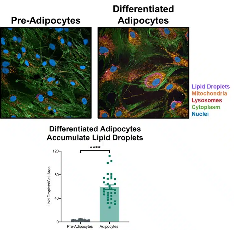
Cell differentiation is marked by transcription, morphological, compositional, and functional changes as cells mature to perform specific physiological roles. Cell painting assays can reveal the morphological and compositional alterations inherent to cell differentiation through high-resolution cell imaging and quantitative image analysis for signatures of differentiation.
Here, pre-adipocytes in culture were treated to stimulate differentiation. Cell painting was performed to image the cell morphology, using labels for the mitochondria, lysosomes, cytoplasm, and nuclei. Additionally, a probe that specifically recognizes lipid droplets was used to interrogate the formation of lipid droplets known to occur in mature adipocytes.
Cell painting visually demonstrates increased lipid droplet accumulation in differentiated cells, and automated quantitation provides enumeration of this trend for statistical analysis.
3D Cell Painting Assays
Spheroid and Organoid Imaging
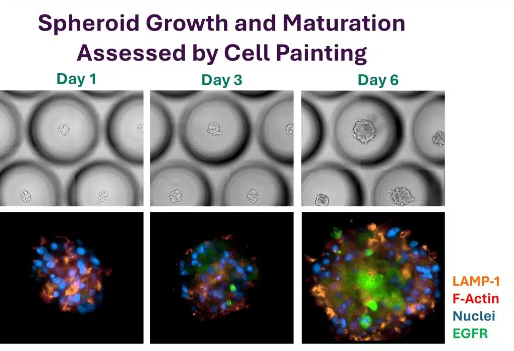
Spheroids and organoids provide relevant models for disease and drug studies. The 3 dimensional (3D) architecture of these cell assemblies can recapitulate whole-tissue dynamics, including cell differentiation and local microenvironments, better than cell culture monolayers. Ichor’s cell painting assays can image 3D cell assemblies through Z-stack assemblies, to assess cell differentiation, receptor or surface marker presentation, drug response or disease state, and overall architecture at distinct Z-planes
Here, a transformed cell line was grown into a spheroid over 6 days, with period imaging of live spheroids by differential interference contrast, and of fixed and labeled spheroids by cell painting. The time course demonstrates spheroid growth over time, and increases EGFR expression and surface presentation as spheroids mature.
Subcellular Compartment Studies
Automated Membrane Masking
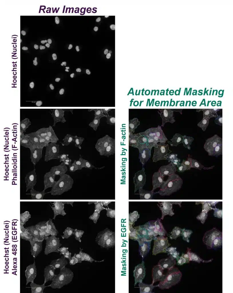
Our cell painting workflows enable automated identification of subcellular compartments and organelles. Software processing algorithms analyze user-input information on fluorescent probes and markers to determine cell nuclei, organelles, membrane borders, and more.
Here, two approaches were compared for their ability to automatically identify cell borders. For both approaches, cell nuclei were identified by Hoechst staining. Then, for the first method, F-actin staining with phalloidin was used for automated determination of cell borders, and a pseudo-colored mask was superimposed on the image to delineate adjacent cell borders. For the second method, Alexa 488-conjugated anti-EGFR was used to stain cell surface EGFR, and automated analysis was again used to superimpose a pseudo-colored mask defining cell borders. Importantly, both methods demonstrated high concurrence of membrane area masking.
This automated recognition of cell features allows for the quantitative analysis of fluorophore intensity within specific subcellular features.
Frequently Asked Questions
Explore our cell painting capabilities below.
Depending on your project, you may receive original grayscale images from individual emission channels, pseudo-colored merged images, and masked images. Ichor also provides raw data from automated calculations, and graphed trends with statistical analysis.
Our instruments perform automated quantitation of probe intensity or area from unbiased field of view sampling. This measurement can be performed for an entire field of view, or for designated regions, like the nucleus or membrane.
Our instruments provide high-resolution imaging of a broad spectrum of fluorophores. Chemical probes and conjugated antibodies are easily incorporated into labeling workflows, and high-performance confocal microscopy technology minimizes signal overlap to enable multiplex cell painting workflows.
Certainly. Our scientists can assemble custom labeling panels to ensure instrument compatibility, with adequate emission discrimination between multiplexed fluorophores.